Jumat, 27 Februari 2009

Joan Rivers Comments on Plastic Surgery


A recent article contains some of Joan Rivers' comments on plastic surgery. Here is an excerpt:

Her collaboration with Valerie Frankel, titled "Men Are Stupid … And They Like Big Boobs: A Woman's Guide to Beauty Through Plastic Surgery," is "really a serious book, but I wrote it funny, as if I'm talking to you — because I'm funny. … If you want to put a chin in, go to the chapter on chins. If you want to raise your breasts, it will tell you every thing you want to know."To research the guide, which offers helpful information on the gamut of cosmetic surgery options, she and her co-writer interviewed 25 plastic surgeons. "They are so boring, these men," she said... In spite of the title, which she claims is something Marilyn Monroe once said to her, Rivers recommends plastic surgery as "something you should do for yourself.""But society is based on looks," she said. "And anyone that says that's not so, is an idiot. If you think big boobs make you look nice, put them in."Men should have work done too, she said. "I hate a turkey neck on anyone."Always up front about her own plastic surgery, which she began in college with her nose being "thinned, not fixed," Rivers added a postscript to anyone who doesn't think they need it. "If you are happy with the way you look, that's wonderful. How lucky you are."

Boring??? The truth hurts sometimes...

Photo credit: prphotos.com

Thanks for reading.

Michigan-based Plastic Surgeon
Anthony Youn, M.D.
:


Selasa, 24 Februari 2009

More Details on Usher's Wife Tameka Foster's Plastic Surgery Complications


More details are emerging regarding the complications suffered by Usher's wife Tameka Foster as she underwent liposuction surgery while in Brazil. Apparently her doctor, Brazilian plastic surgeon Dr. Silvio Sterman, planned to perform liposuction on her while she was only two months postpartum. After being placed under general anesthesia she suffered a cardiac arrest and was revived via CPR. She was then placed into a drug-induced coma in order to run tests and make sure she was stabilized. She has now been discharged from the hospital and is said to be "doing very well."

I do not perform any cosmetic surgery on women who are less than 6 months postpartum. There is no scientific studies which determine that 6 months is a mandatory time frame, but I believe the changes that occur in a woman's body (both physical and hormonal) can persist for months after a baby is born. Liposuction just 2 months after giving birth? In my opinion that is too soon.

Source

Thanks for reading.
Michigan-based Plastic Surgeon
Anthony Youn, M.D.
:

Minggu, 22 Februari 2009

Mickey Rourke Admits to Plastic Surgery by Hack Surgeon


According to Monsters and Critics, Oscar-nominated actor Mickey Rourke has admitted to having plastic surgery by a hack plastic surgeon. He reportedly stated,

"Most of it was to mend the mess of my face because of the boxing, but I went to the wrong guy to put my face back together... I had my nose broken twice. I had five operations on my nose and one on a smashed cheekbone... I had to have cartilage taken from my ear to rebuild my nose and a couple of operations to scrape out the cartilage because the scar tissue wasn't healing properly. That was one of the most painful operations, but the worst was hemorrhoids."

It's funny that he admits to having hemorrhoids but doesn't admit having a facelift, which he appears to have had. For my opinions on his previous surgery, click here.

I hope he wins the Oscar tonight. I saw his performance in The Wrestler and think it's amazing. He really deserves it. I'd much rather see him win it instead of that smug Sean Penn.

Photo credit: prphotos.com

Thanks for reading.
Michigan-based Plastic Surgeon
Anthony Youn, M.D.
:


Sabtu, 21 Februari 2009

Alloplastic Materials

Introduction
Advances in medical technology have allowed plastic surgeons to utilize synthetic materials as an alternative to autologous tissues when performing many of today’s aesthetic and reconstructive surgeries. Although autologous materials are generally preferred, synthetic materials provide several advantages over tissues obtained from the patient:

  • Not resorbed over time (unless they are designed to do so)
  • Do not require a second surgical donor site
  • Provide more material than can often be obtained from the patient
  • Can be custom-tailored to the individual patient
  • Reduce operating time since graft harvesting is not performed

Because of the many benefits to using alloplastic materials, there is currently a strong interest in developing the ideal implant material which would possess the following characteristics: it should (1) be chemically inert; (2) be incapable of producing hypersensitivity or a foreign body reaction; (3) be easily contoured; (4) retain stable shape over time (except when desired); (5) be noncarcinogenic; (6) become ingrown or replaced by living tissue; (7) be easy to remove and sterilize; and (8) not interfere with radiographic imaging. Despite much effort and ingenuity, creation of the ideal implant material has yet to be accomplished. However, various alloplastic materials are being used today in plastic and reconstructive procedures, and many of them have proven quite promising.

Pre- and Intraoperative Considerations
The vascularity of the recipient site and the ability to provide sufficient soft tissue coverage of the implant must be assessed preoperatively. Decreased vascularity secondary to scar tissue (from previous surgeries) or radiation impairs the establishment of normal fibrovascular tissue encapsulation and may interfere with the normal inflammatory response if the implant were to become infected.

In order to prevent implant exposure or extrusion, soft tissue coverage over an implant should be as thick as possible. The size of the implant should be comparable to that of the tissue pocket or wound cavity in order to avoid tension of the overlying soft tissue, and the implant should be fixated to a stable adjacent structure to prevent migration of the implant. All patients should receive perioperative intravenous antibiotics followed by a postoperative oral course, although the optimal antibiotic choice and duration have yet to be determined for most implants. What is clear is that intraoperative handling of the implant should be minimized in order to prevent bacterial transmission, and strict adherence to sterile technique is essential.

Classification of synthetic materials used in plastic and reconstructive surgery

Silicone-based materials:

BioPlastique

Injectable silicone

Silastic sheets

Silicone

Silicone gel

Polytetrafluoroethylene:

Gore-Tex

Proplast I and II

Teflon

High density polyethylene:

Medpor

Polymer mesh:

Dacron (Mersilene)

Dexon

Prolene

Supramid Vicryl

Biological glasses:

Bioactive glasses (Bioglass)

Glass ionomer

Tissue adhesives:

Cyanoacrylate

Acrylics:

HTR Polymer

Methylmethacrylate

Choice of Alloplastic Material
The type of procedure as well as the size and character of the defect being augmented often dictate the type of implant material. In the preantibiotic era, inert materials such as gold, silver, platinum and paraffin were used with little success and were quickly abandoned. Currently, there are numerous implantable materials being used today (Table 14.1). These materials are used in a wide range of procedures, such as aesthetic procedures, craniofacial surgery, maxillofacial trauma, breast reconstruction and hand surgery. Table 14.2 lists the common uses for the various allopastic implants.

Silicone
Silicone-based prosthetics have been used as medical implants since the 1950s due to their chemically inert nature, resistance to degradation, and lack of significant allergic reactions. Silicone is useful for a variety of aesthetic surgeries, complex contouring and reconstructive procedures. Silicone comes in the form of silicone gels, silicone rubber or solid silicone implants. Silicone gels can provide a more natural feel, as seen with breast implants, but the risk of rupture requiring capsulectomy is a distinct disadvantage. The use of silicone gel has been surrounded by controversy related to concerns about migration, toxicity and an unproven association with systemic disease, leading to restriction of the use of silicone gel implants by the FDA in 1992. This ban was recently lifted after an extensive unbiased review by the Institutes of Medicine. Silicone rubber is used for tissue expanders, the outer shell of both saline-filled and silicone gel-filled breast implants, and as an onlay material for the augmentation of the bony skeleton and soft tissues. However, silicone rubbers are relatively weak and tend to tear, leading to implant failure. Solid silicone implants are commonly used for chin and malar augmentation, and have been used in nasal, chest and calf augmentation, as well as in joint replacement and tendon reconstruction.

A list of the procedures that commonly employ allopastic materials

Procedures

Materials Used

Cranioplasty and forehead augmentation

Glass ionomer and bioactive glass Hard-Tissue-Replacement (HTR) polymer

Methylmethacrylate

Medpor

Poly(L-lactide) and polyglycolic acid plates and screws Silicone

Anterior mandibular augmentation

Medpor Polyamide

mesh

Silicone

Mandibular body and angle augmentation

Glass ionomer and bioactive glass

Medpor

Methylmethacrylate

Poly(L-lactide) and polyglycolic acid plates and screws

Malar and maxillary reconstruction

Glass ionomers

Gore-Tex

Medpor

Methylmethacrylate

Polyamide mesh

Silicone

Teflon

Zygomatic reconstruction

Glass ionomers

Medpor

Gore-Tex

Poly(L-lactide) and polyglycolic acid plates and screws

Silicone

Orbital reconstruction

Gore-Tex

HTR Polymer

Medpor

Poly(L-lactide) Silicone

Teflon

Ear reconstruction

Medpor

Silicone

Tendon repair

Gore-Tex

Cyanoacrylate

Soft tissue augmentation

BioPlastique

Gore-Tex

Breast augmentation and tissue expansion

Silicone (saline or silicone gel filled)

Wound repair and scar revision

Cyanoacrylate

Silastic sheets

Chest and abdominal wall reconstructions

Dacron mesh

Gore-Tex

Prolene mesh

Vicryl mesh

Nasal augmentation

Gore-Tex

Polyamide mesh

Silicone

Because silicone is not porous, tissue ingrowth does not occur. A fibrous capsule forms around the implant that is relatively avascular and can contract which may lead to implant migration. This avascular capsule is a potential space for infection and in the setting of infection may require removal of the implant.

BioPlastique
BioPlastique® is a nonbiodegradable, relatively inert injectable liquid used for soft tissue augmentation. The textured surface of the particles allows for tissue ingrowth, and the particle size is large enough to prevent engulfment by macrophages but small enough to become encapsulated within 3 to 6 weeks. Studies on the use of BioPlastique demonstrate good-to-excellent results in augmenting small defects on the dorsal nose, malar area, cheeks and chin with no adverse immunologic reactions. Although the clinical results with Bioplastique have been encouraging, it is not FDA approved at this time.

Polymethylmethacrylate
Polymethylmethacrylate (PMMA) is an acrylic polymer used as a bone substitute in plastic surgery and neurosurgery. PMMA is radiolucent, extremely durable and completely biocompatible, making it a widely used material for cranial bone reconstruction-alone or in combination with wire or mesh reinforcement. When powdered granules of methylmethacrylate polymer are mixed with methylmethacrylate liquid monomer, a moldable dough forms as the monomer polymerizes and hardens in about ten minutes. Near the end of the polymerization process, an exothermic reaction occurs that can potentially damage the local tissues, the major complication associated with the use of PMMA. This can be avoided by continually irrigating the implant bed with cool saline during the polymerization. A rare, but serious complication is the inadvertent entry of the PMMA into the venous or arterial systems. If this occurs it can cause complete heart block, cardiac arrest and other arrhythmias. This complication is most often seen during orthopedic procedures where PMMA is used for joint replacements or fracture repair. Hard-tissue-replacement (HTR) polymer is a porous form of PMMA that allows for fibrous ingrowth, leading to an implant that is nonresorbable and very stable. Applications for HTR include chin and malar augmentation, with potential for additional uses in craniofacial reconstruction.

Polyester (Dacron®, Mersilene®)
Polyethylene terephthalate (Dacron) is a biocompatible, flexible, nonabsorbable polymer that is used as a suture material, as a prosthetic material for arterial replacement, and as a mesh (Mersilene) in abdominal and chest wall reconstruction. Its use has also been described for chin and nasal augmentation. Biodegradable Polyester (Polyglycolic Acid, Poly-L-lactic Acid) Polyglycolic acid (PGA) and Poly(L-lactide) (PLLA) are polymers that are degraded in the body at physiologic pH over the course of weeks to months. These resorbable polymers are available as mesh sheets for body wall reconstruction and as rods for the internal fixation of fractures and osteotomies. They have also been fashioned into resorbable miniplates and screws for the fixation of bones of the craniofacial skeleton. Although they do not appear to have any cytotoxic effects, they do provoke an inflammatory or foreign body response after implantation.

Polyamide Mesh (Supramid®, Nylamid®)
Polyamide mesh is a woven, polymer mesh implant that is biocompatible, can be easily shaped and sutured, allows for fibrous tissue ingrowth, and has been used for the repair of orbital floor defects. It seems to be well tolerated and has a low rate of extrusion, even in areas of thin skin such as the nasal dorsum. However, polyamides do undergo resorption and induce an inflammatory response, making their use in facial augmentation and reconstruction somewhat limited.

Porous Polyethylene (Medpor®)
Medpor is a high-density, porous polyethylene implant used frequently in facial surgery because it is nonantigenic, nonallergenic, nonresorbable, highly stable and easy to fixate. In addition, Medpor is available in a wide variety of preformed shapes for its use as a malar, chin, nasal, orbital rim, orbital floor and cranial implant, as well as an auricular framework in postburn ear reconstruction. Overall, complications of Medpor, such as exposure or infection, are rare.

Polytetrafluoroethylene (Teflon®, Gore-Tex®, Proplast®)
Polytetrafluoroethylene (PTFE) is an inert and highly biocompatible polymer that is extremely useful in soft tissue augmentation but has limited use in bony repair due to its low tensile and compressive strength. Teflon, the first PTFE graft to be used in plastic surgery, is a chemically inert polymer used for soft tissue augmentation in the past, but the main application for Teflon has been orbital floor reconstruction. Gore-Tex is a pliable, durable, inert, biocompatible material that has some tissue ingrowth, little inflammatory reaction and almost no encapsulation. In addition to being used in abdominal fascial reconstruction, chest wall reconstruction and soft tissue reconstruction, Gore-Tex has also been utilized for lip, nasal, chin and malar augmentation. It has also been utilized for the treatment of nasolabial and glabellar creases. Proplast I is a highly porous, black graphite/PTFE composite with a spongy consistency. Because Proplast I led to discoloration of the surrounding soft tissues when implanted, Proplast II-a more rigid, white PTFE/alumina compound-was developed as an alternative. Proplast, with a wide variety of applications including the reconstruction of the chin, zygoma, orbital rim, maxilla, mandible, skull and rib cage, was originally regarded favorably. However, reports of biomechanical failure, intense inflammation, infection and extrusion related to the Proplast temporomandibular joint implant, led to the removal of all Proplast implants from American markets by the FDA in 1990.

Calcium Phosphate Ceramics
Calcium phosphate implants have been available as bone replacement/augmentation materials for 20 years. The primary calcium phosphates in clinical use are hydroxyapatite and tricalcium phosphate. These materials are osteoconductive (providing a scaffold for bone ingrowth) thus allowing for integration into the recipient site after placement. As a result, calcium phosphates are very well tolerated with essentially no inflammatory response, minimal fibrous encapsulation, and no adverse effects on local bone mineralization.

Metals
Metals have been used for the past 35 years for skull reconstruction and repair, in addition to reconstruction of craniofacial and upper extremity skeletal injuries. Stainless steel, cobalt-chromium (vitallium), pure titanium and titanium alloys are the principal metals currently available. Characteristics of a desirable metal implant include biocompatibility, strength, resistance to corrosion and imaging transparency.

Postoperative Considerations
Although numerous potential complications may occur with any implant-related procedure (e.g., migration, extrusion, palpability), the one common denominator shared by all alloplastic implants is their inherent risk of infection. The majority of postoperative infections appear within weeks to months after the initial surgery. Low-grade infections manifested only by fevers and signs of mild cellulitis are managed by intravenous antibiotics. More serious infections involving wound breakdown, implant exposure, gross purulence or systemic spread of the infection require prompt removal of the implant as antibiotics and drainage alone are usually insufficient. Reimplantation should not be performed for at least 3 to 6 months to allow for complete treatment and resolution of both the infection and the inflammation in the surrounding tissues. Several studies suggest that smooth, nonporous, nonresorbable implants have lower rates of infection, but it remains to be seen whether any true infectious risk differences exist among the various alloplastic implant materials available today.

Pearls and Pitfalls

  • Incisions should be placed as far as possible from the final position of the implant. This will decrease the risk of implant exposure or extrusion in the setting of a minor wound infection.
  • The implant should be covered with as much soft tissue as possible. The pocket should be of adequate size; too large and the implant will shift position, too small and the implant will be at risk for extrusion due to tension on the closure.
  • Whenever possible, always try and close a second layer of tissue between the skin and implant. This is critical if the implant lies directly beneath the incision.
  • Implants with sharp corners should be smoothed down, since sharp edges can erode through the skin with time.
  • The implant should be touched as little as possible. Clean, powder-free gloves should be worn and instruments should be used to handle the implant whenever feasible. The risk of infection and abnormal capsule formation is increased by the presence of any bacteria or foreign material on the implant.
  • Do not use an implant composed of a rigid material to replace soft, pliable tissue.
  • Keep an organized registry of all alloplastic implants in the event that the device fails or has to be removed. Give the patient a copy of the device name, model, manufacturer and serial number, in case failure occurs in the care of another physician.

Jumat, 20 Februari 2009

Cosmetic Surgeon Goes After Joan Rivers to Get Publicity


A general/vascular/cosmetic surgeon in California has just sent out a press release attacking Joan Rivers' guide to plastic surgery. In his press release he states,

"While Joan Rivers certainly has a right to hawk her beliefs about plastic surgery, her book and the half dozen reality shows that focus on plastic surgery undermine the credibility of the beauty enhancement business."

Who is this guy to talk? He's a general and vascular surgeon who practices cosmetic surgery! To me it looks like a blatant attempt to hop on Ms. Rivers' coattails and get a little media coverage. I find Joan's book well-written, humorous, and chock-full of useful information for anyone considering plastic surgery. Maybe he didn't like the chapter where she encourages everyone to seek a surgeon certified by the American Board of Plastic Surgery.

Click here for the press release. To purchase a copy of Joan's book, click below.


Disclaimer: I am a contributor to Joan Rivers' book, but do not receive any payment or royalties for the sale of her book.

Thanks for reading.
Michigan-based Plastic Surgeon
Anthony Youn, M.D.
:

Rabu, 18 Februari 2009

Tom Cruise Visits Brazilian Plastic Surgeon?


Page Six of the New York Post is reporting that Tom Cruise visited prominent Brazilian plastic surgeon Dr. Ivo Pitanguy while recently in Rio De Janeiro. He took a side trip to visit Dr. Pitanguy on the doctor's private island which features its own landing strip and exotic animals.

Did Tom have some work done while visiting there? He does look awfully youthful lately, but I bet the most he may have had done is a chemical peel and some minor facial treatments.

Wow. I wish I had a private island...

Photo credit: prphotos.com

Thanks for reading.
Michigan-based Plastic Surgeon
Anthony Youn, M.D.
:

Senin, 16 Februari 2009

Celebrity Eyebrow Makeovers on MSNBC.com


MSNBC.com has an interesting slideshow in the Top 12 Celebrity Eyebrow Makeovers. Some are huge improvements while others are barely noticeable. You can check it out here.

It reminds me of a photo I show some patients. I once had a woman in her 30's see me in consultation for a browlift. After looking at her for about two seconds I figured out that she didn't need a brow lift, but a brow wax. Fifteen minutes (and twenty dollars) later she looked a heck of a lot better, as her Andy Rooney bushy brows were shaped into nice, defined arches. I saved her about $3500 and she left the office happy and surgery-free!

Thanks for reading.
Michigan-based Plastic Surgeon
Anthony Youn, M.D.
:

Sabtu, 14 Februari 2009

CNN on the OctoMom / Angelina Jolie resemblance


CNN's Jeanne Moos reports in this video on the resemblance between the OctoMom and Angelina Jolie. The more I hear about the OctoMom the weirder she seems. Rhinoplasty and Lip augmentation - Looks like it. Botox and blepharoplasty - Maybe.

Thanks for reading.
Michigan-based Plastic Surgeon
Anthony Youn, M.D.
:

Jumat, 13 Februari 2009

Does Michael Jackson Have An MRSA Infection?


Several internet sources are reporting that Michael Jackson may be fighting a severe MRSA infection on the face. These sources state that he is on IV Antibiotics for treatment of this multiple drug-resistant bacteria, and that it may have originated from nasal surgery. Could this be true?

There doesn't appear to be any word from Michael's spokesperson yet, which may be a bad sign. MRSA is a type of Staph aureus bacteria which is often found inside the nose of "carriers." Although usually asymptomatic, in rare cases it can cause severe infections which can even destroy tissue. There are very few drugs which are effective against MRSA, although a handful do exist. The photos of Michael show a very limited amount of the skin of his face (since it is covered by a surgical mask), but the skin that is shown does appear red and possibly inflamed. For his sake, hopefully the rumors are false and he remains in good health. I have never seen a serious MRSA infection of the face (although I have seen it elsewhere), but I can imagine it can be pretty scary.

Source: The Sun

Photo credit: prphotos.com

Thanks for reading.
Michigan-based Plastic Surgeon
Anthony Youn, M.D.
:

Rabu, 11 Februari 2009

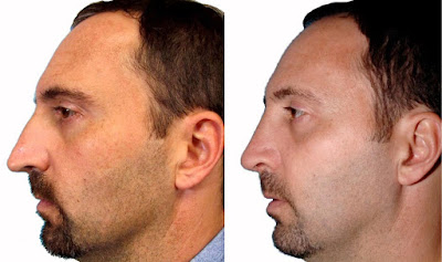
Nose Job Before After

Nose Job Before After

Nose Job Before After

Nose Job Before After


Nose Job Before After


Nose Job Before After


Nose Job Before After


Nose Job Before After


Nose Job Before After


Nose Job Before After


Nose Job Before After


Nose Job Before After

Tameka Foster- Usher's Wife - Update on Plastic Surgery Complications


There are a few news stories that are reporting the current status of Usher's wife, Tameka Foster, who had complications while undergoing plastic surgery in Brazil. They all state that she is in stable condition, which is good. Details are still sketchy, but they include a possible cardiac and respiratory arrest during liposuction surgery. The anesthesia has been implicated as a possible cause, and she may have been induced into a coma for 24 hours.

Hopefully she hasn't suffered anoxic brain injury or another very serious complication. This story continues to develop, and I hope she comes out of it ok.

Story links: azcentral.com, stuff.co.nz
Photo credit: prphotos.com

Thanks for reading.
Michigan-based Plastic Surgeon
Anthony Youn, M.D.
:

Introducing SlimLipo™ — The Kinder, Gentler Liposuction

As mentioned in a previous post, I was smitten by the new laser-assisted liposuction technologies that I saw at the recent American Society of Plastic Surgeon annual meeting in Chicago. So I ordered the SlimLipo system from Palomar, and have been offering it here in my Duxbury plastic surgery practice since December. The results have been great and as expected — the new laser system actually

Senin, 09 Februari 2009

Dr. Youn on the Rachael Ray Show


Here is a link to a clip of me on the Rachael Ray Show that aired today. Rachael and I discussed some of the newest trends in plastic surgery.
Click here for the clip!

Thank you to everyone at the Rachael Ray Show for inviting me!

Photo credit: prphotos.com

Thanks for reading.
Michigan-based Plastic Surgeon
Anthony Youn, M.D.
:

Now Offering: Latisse for Eyelashes

Here's more on Dr. Hamori's latest offering in her Duxbury, Massachusetts practice: Latisse for longer, thicker lashes, which she mentioned in her previous post.Recently approved by the FDA for treatment of "hypotrichosis" of the eyelashes — another name for having inadequate or not enough eyelashes — Latisse™ enhances eyelash prominence as measured by increases in length, thickness and darkness

Minggu, 08 Februari 2009

Usher's Wife - Complications from Plastic Surgery in Brazil


Sources are reporting that Usher's wife, Tameka Foster, has suffered major complications from plastic surgery performed in Brazil. She gave birth only two months ago. A neurosurgeon has been flown from the U.S. to Brazil to help with the situation.

Details are still sketchy, but this case appears to highlight the dangers of having any medical procedure performed in another country. Plastic surgeons certified by the American Board of Plastic Surgery adhere to strict education, experience, safety, and ethical standards which just aren't comparable to care given in other countries. The American Society of Plastic Surgeons has issued a cautionary statement to the public regarding 'medical tourism.' Also, your health insurance will probably not cover medical care given in another country and the complications that can ensue.

Why would a neurosurgeon be flown to Brazil for a plastic surgery-related complication? I'll give some of my thoughts in tomorrow's post.

Photo credit: prphotos.com

Thanks for reading.
Michigan-based Plastic Surgeon
Anthony Youn, M.D.
:

Heidi Montag and Spencer Pratt


Hollywood tuna has a bunch of photos of Heidi and Spencer doing a photo shoot in the pouring rain. Who does this?They look like complete tools. At least Heidi had some good plastic surgery. Hopefully these two will go away sometime soon.

Click here for my other posts on Heidi Montag's good (and not so good) plastic surgery.

Thanks for reading.
Michigan-based Plastic Surgeon
Anthony Youn, M.D.
:

Sabtu, 07 Februari 2009

Microvascular Surgical Technique and Methods of Flap Monitoring

Introduction

The hand is capable of coordinated activity finer than the eye can direct. With the aid of magnification, the true capability of the hand can be exploited. As a tool for the plastic surgeon, microsurgery has allowed reconstructions that were simply not possible before. However, microvascular free tissue transfer is not a technique for the occasional microsurgeon. The catastrophic complication of flap failure looms over every microsurgical case; therefore, expertise in the execution of a free flap as well as its postoperative surveillance is key to a successful outcome.

Experience has shown that flap loss is a preventable complication and that elective microsurgery should have a failure rate of less than 2%. Most cases of flap loss are technical in nature. The fault may lie in the choice of flap, the harvest of the flap, preparation of donor vessels, insetting of pedicle or microsurgical technique. In general, it is best to think of all possible errors as additive in the process of thrombosis. Failure will occur if the procoagulatory factors outweigh the intrinsic ability of the vessels, in particular intact and uninjured intima, to prevent clot formation.

Flap Choice

The first step for success in microsurgery is flap choice. The specifics of different flaps are discussed in subsequent chapters. The most important determining factors for flap choice should be the surgeon’s experience and the goals of reconstruction.

In general, each surgeon should identify at least four flaps they feel comfortable with. These flaps should include a bulky muscle flap, a bulky fasciocutaneous flap, a thin fasciocutaneous flap, and a bone flap. With this armamentarium, the reconstructive surgeon will have tools that can be applied to most situations. By limiting himself to a small number of flaps, more experience can be obtained with each one. This increased experience translates to increased success. It is not advantageous to explore every novel flap that is reported, as this dilutes the experience and increases the chance of failure. With increasing experience with each flap comes increasing success and a lower failure rate.

This does not imply that specific flaps may not be beneficial over others in certain situations. There is no doubt that the donor properties of a latissimus dorsi flap differ from those of the gracilis flap and that each may be a better choice for a specific patient. However, the patient is best served with successful reconstruction. If there is significant benefit in a flap where the surgeon has no experience, the surgeon should consider referral or should seek additional training in order to add that flap to his or her armamentarium. This may include time in a cadaver lab or observing a surgeon with a particular skill.

Having mastered the tools of reconstruction, the surgeon should judiciously consider the requirements for reconstruction. Bulky muscle flaps are best for contaminated defects and bony injuries with high risk for infection. Thick fasciocutaneous flaps are useful for contour and shape reconstruction. Thin fasciocutaneous flaps provide stable, noncontracting coverage. Bone flaps provide structural integrity.

,

Specific aspects of each flap harvest are discussed elsewhere in this book. Certain principles, however, hold true despite the flap chosen. While harvesting a flap, the pedicle should be carefully dissected with as much length as possible. It is important not to limit the pedicle length to the anticipated need, but to harvest the maximum that can safely be obtained. It is much more advantageous to discard unneeded length than to find oneself requiring more pedicle length. Vein grafts should be avoided unless absolutely necessary.

While harvesting the flap and dissecting the pedicle, the most common mistake is damaging the vessels. Forceps should only touch the adventia and never purchase the vessel as the intimal layer is extremely fragile and easily fractured or crushed by manipulation. Any grasping of the vessels will cause damage to the intima which increases the likelihood of clot formation. This intimal injury leads to platelet deposition and thrombosis as the injured endothelial cell layer loses its natural thrombolytic properties.

Division of the pedicle should be reserved until the last possible moment. Prior to division, the donor vessels should be dissected, isolated, prepared and positioned for the anastomosis. It is helpful to mark the vessels in their natural state to assure that they are not twisted when transferred to the recipient site. Prior to division, the artery should be occluded first, followed by the vein. This will avoid excess blood pooling in the flap. Immediately after the flap is removed, one can consider cooling the flap with chilled saline as this decreases the metabolic activity of the tissue and allows the luxury of a longer ischemic time.

There is seldom a need to separate the artery and vein within the pedicle for anything more than a minimal distance. The only exception is the case where the recipient vessels are not paired. The vessels should not be skeletonized until they are brought to the recipient site and carefully prepared under the microscope. Any branches within 2 mm of the anastomosis are best sutured closed with microtechnique to avoid blood pooling near the anastomosis.

Preparation of Recipient Site

Preparing the recipient site mirrors the harvest of the flap. Vessels should be chosen that are simple to use and of the largest caliber available. They should be expendable when possible and have sufficient length. Again, care should be taken in the preparation of the vessels. They should not be extensively manipulated or injured. They should only be skeletonized for 2-3 mm around the anastomotic site, and this should be done under the microscope.

Microsurgical Technique

The anastomosis can be done in several fashions. These include end-to-end or end-to-side. They can be performed by multiple suture techniques or with coupling devices. The general philosophy is to gain experience with two or three techniques and apply those techniques to different situations. With careful planning, preparation, and mobilization of both the pedicle and recipient vessels, this is generally possible.

General principles of proper microsurgical technique are:

  1. Pass sutures perpendicularly through the adventitia into the intima.
  2. Avoid grasping or manipulating the intima.
  3. Avoid multiple suture passes.
  4. Avoid torquing the needle in the vessel; grasp and regrasp the needle in order to pass it through the vessel following the curve of the needle perfectly.
  5. Dilate and visualize the inside of the vessels with heparinized saline irrigation on an ocular anterior chamber needle.
  6. Use polished vessel dilating forceps to gently open spasmodic vessels or for vessel expansion.
  7. Leave long tails on the sutures for manipulation and visualization.
  8. Perform both anastomoses prior to reperfusion.
  9. Release clamps on the vein first.
  10. Inspect the anastomosis using the long suture tails as handles.
  11. Place additional sutures in gaps with pulsatile or pressurized bleeding.
  12. Avoid the temptation to place excess sutures in cases of mild oozing of blood from the anastomosis.
  13. Apply warm saline to the flap and papaverine to the anastomosis after reperfusion to dilate the vessels and relax spasm.

Anastomotic Techniques

End-to-End

The end-to-end anastomosis is the simplest and the most reliable method. There are several techniques of suture placement including the 180˚-180˚ and triangulation methods. The easiest is probably the 180˚-180˚ technique. This can be applied to any situation and is probably the best technique for size-mismatched vessels.

Important points to remember are:

  1. The vessels must not be twisted prior to placement in the double clamp holder. This can be ensured by inking one surface of the pedicle and recipient vessels prior to their division or dissection.
  2. The first two sutures are placed at opposite poles of the vessels.
  3. The third suture is placed midway between the poles.
  4. In most cases, the next sutures bisect the gap though on occasion, two sutures will be needed in the gap.
  5. Once the anterior wall is complete, twist the entire double clamp to show the backwall.
  6. Visually inspect every suture of the anterior wall from the posterior view to assure that they are evenly spaced and have not purchased the back wall of the vessel.
  7. Place another bisecting suture midway between the poles on the back wall.
  8. All remaining sutures can be placed and left long (not tied).
  9. Dilate the vessel with saline when tying the back wall to assure that there is no purchase of the anterior wall.

End-to-Side

The end-to-side technique is occasionally necessary. For example, it is used in the leg when there is only one vessel available or for an anastomosis in the head and neck (for example, to the internal jugular vein). Principles are:

  1. The pedicle vessels should enter the recipient vessel at a gentle angle.
  2. Perform a limited arterioectomy, removing a small window of vessel.
  3. Place heel and toe sutures first.
  4. Initially close the heel.
  5. Follow with closure of the toe.

Coupling Devices

Coupling devices are useful for veins or thin-walled arteries. They save some time in the anastomosis. They, however, are not a panacea. The major time consumption in a microsurgical case is not the anastomosis, but the set up and preparation. If coupling devices are used, the set up and preparation time remain the same. Principles of gentle handling of vessels are still required as is avoidance of damage to the intima. Overall, the devices appear to have a place in the venous anastomosis, where they can also act as a stent, or in cases with significant size mismatch. Points to consider are:

  1. Use the largest size coupler that will comfortably fit (range 2-3.5 mm).
  2. Draping of the vessel over the spikes is performed by one surgeon while the other maintains the engagement of the spike as the vessel is seated.
  3. Seat the vessel 180˚ apart to assure even spacing on the coupler.
  4. Avoid grasping the intima of the vessel as it is draped over the spikes.
  5. Assure that the coupling device is closed and guide it off of the coupling applier.

Draping of the Pedicle

After the anastomosis is complete and the flap is successfully revascularized, it is not uncommon for significant problems to arise. Kinking or unnatural curvature of the pedicle will certainly cause thrombosis. In fact, any turbulent, nonlaminar flow increases the likelihood of thrombosis and flap loss. The pedicle should be carefully draped. Gelfoam sponge or Alloderm can be used to help maintain the proper position of the pedicle.

Closure

A sound closure technique is again crucial for success. Both the flap and pedicle can be compressed by a tight closure. Anticipation of this is critical, as well planned incisions will allow closure after the edema of these long cases has set in. If there is any question, the liberal use of skin grafts to allow tensionless closure is recommended. The anastomosis should never be situated immediately under a suture line.

Monitoring

There is no “perfect” monitoring technique. Despite numerous ingenious techniques and improvements in technology, the ideal monitoring technique should be the one that surgeons and ancillary staff at a particular hospital are most familiar with and meet the restraints (budgetary or manpower) of the institution. What is ideal at one institution may not be practical at another. What is clear over many years of clinical experience, although this remains to be formally proven, is that the presence of dedicated staff in a dedicated unit stands the best chance of picking up problems earlier. The impetus to closely monitor a flap comes from the enormous investment undertaken on the part of the patient as well as the surgeon regarding microvascular free tissue transfer. The utility of postoperative flap surveillance has been proven, with an increase in the salvage rate of the failing flap from 33% to about 70% in some series.

The clinical exam is useful when performed by the experienced clinician. The transition of a healthy, plump flap or vibrant replanted digit to cold, flat, lifeless tissue can proceed via either arterial occlusion or venous congestion. These characteristics are useful in deciding whether to explore a flap or perhaps treat with leech therapy. Although it is the least technologically-based, much information can be gleaned from a thorough physical exam. Turgor can indicate the state of arterial inflow or venous outflow. Like a balloon, the flap or digit will inevitably declare itself if it has arterial insufficiency or venous congestion. Bleeding can be useful, as the qualitative and quantitative flow in response to pinpricks or rubbing of wound edges can declare the state of circulatory flow to the flap. In particular, a congested flap may bleed briskly, but the blood will appear dark and unoxygenated. The blood flow of a flap with compromised arterial inflow will be weak or absent. A caution regarding the pinprick test is that it is useful for evaluating a flap, but will occasionally cause trauma leading to vasospasm or hematoma in the confined space of a finger.

It is possible to monitor free flaps with a temperature probe. This method consists of placing surface temperature probes on the skin of the free flap and comparing them to probes placed on neighboring native skin. The probes are attached to a temperature monitor that will give off an alarm if there is a difference in temperature between the two sites greater than the specified amount (typically, 2-3˚C). Although appealing, there are limitations to the use of temperature probes, as the readings may be affected by regional changes in blood flow that are not secondary to flap flow disturbances.

Doppler ultrasonography is perhaps the most widely used monitoring tool. Two permutations exist. The first is the external Doppler. A recent innovation is the implantable internal Doppler. This tool permits monitoring of the segment of artery and vein a short distance downstream of the anastomosis. Its use has obviated the need for an external sentinel skin segment, and is ideally suited for buried anastomosis (e.g., jejunal free flaps in the head and neck, or vascularized bone transfers). These techniques are extremely useful; however, complications such as probe dislodgement and the occasional monitoring of an adjacent vessel that is not the pedicle can result.

In replants, the pulse oximeter is extremely useful. Some centers have reported success with fluorescein infusion and fluorescent lamp observation. This technique is not as useful in pigmented skin. Other techniques that at this time must be considered experimental include pH monitoring, duplex ultrasound, photoplethysmography, reflection photometry and radioisotope studies. None of these are currently widely used.

Pearls and Pitfalls

Although the microsurgical trainee may be eager to execute a large variety of occasionally exotic flaps, it is much more important to master a limited number of flaps and apply these flaps to different defects throughout the body. The principles outlined in this chapter serve as the basis to successfully execute any type of microsurgical transfer the plastic surgeon will encounter, even unusual flaps. In summary, it is essential to:

  1. Sharpen microsurgical skills in the lab.
  2. Handle the vessels gently.
  3. Place significant attention on closure and pedicle position.
  4. Familiarize oneself with one or two monitoring techniques. This will maximize salvage of the inevitable free flap failure.

The most important indicator of a problem with the free-flap is a change in the

clinical exam. This necessitates that the flap be seen as soon as possible by a surgeon who has been actively managing the patient.

Popular Posts